
Soft X-ray Emission SpectrometerSXES. The D8 ADVANCE is the benchmark when it comes to extracting structural information from X-Ray Powder Diffraction including Rietveld TOPAS analysis.

Field Emission Scanning Electron Microscope Fe Sem Hitachi S4700 Fe Sem Is A Powerful To Scanning Electron Microscope Electron Microscope Digital Processing
SNOM Chapter 324 has been used to irradiate protein films on silicon and to obtain their mid-IR spectra with a spatial resolution of about 10 nm obtained through the near-field opticsFirst the IR spectra of individual ferritin molecules and those of.

. Life Science Electron Microscopy Lab. A light microscope has a low resolving power 025µm to 03µm while the electron microscope has a resolution power about 250 times higher than the light microscope at about 0001µm. SU5000 combines Schottky emission electron source and out-lens objective lens for high resolution imaging and diverse analyses of samples with various sizes and compositions.
JCM-7000 NeoScope Benchtop SEM. The Jeol field emission scanning electron microscope is a versatile high-resolution scanning electron microscope. We present a protocol for building a scanning light-field microscope with digital adaptive optics as an add-on to a standard wide-field microscope to achieve long-term high-speed intravital.
Life Science Electron Microscopy Lab. High Resolution large chamber FE SEM. Wavelength Dispersive X-ray Fluorescence WD-XRF LIQUID CHROMATOGRAPHY MASS SPECTROMETER LC-MS-MS.
Macro to Nano -- Full Scale Scanning Electron Microscope Solutions. The first Scanning Electron Microscope was initially made by Mafred von Ardenne in 1937 with an aim to surpass the transmission electron Microscope. The SEMView8000 Scanning Electron Microscope SEM universal operators console has been installed at a RDQC manufacturing laboratory.
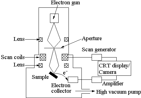
He also aimed at reducing the problems of chromatic aberrations images produced by the. Adam West in Interface Science and Technology 2018. A field-emission cathode in the electron gun of a scanning electron microscope provides narrower probing beams at low as well as high electron energy resulting in both improved spatial resolution and minimized sample charging and damage.
The 300 kV Themis Z is a FEG Scanning Transmission Electron Microscope STEM with a high-tension voltage range of 60-300 kV. TRANSMISSION ELECTRON MICROSCOPE TEM WITH CCD CAMERA. Similarly a light microscope has a magnification of 500X to 1500x while the electron microscope has a much higher magnification of 100000X to 300000X.
Gather-X JED Series Dry SD Windowless EDS. The small tip radius improves emission and focusing ability. Scanning Electron Microscopes.
The first electron microscope EM observation of an individual atom was made by Crewe Wall and Langmore in 1970 using a scanning electron microscope equipped with an early field emission gun. The NEW Thermo ScientificTM Phenom Pharos is the only tabletop SEM with a field emission FEG source. Nanoscience Instruments is a proud partner of Thermo Scientific featuring the worlds best-selling Scanning Electron Microscope.
He used high-resolution power to scan a small raster using a beam of electrons that were focused on the raster. Ultrahigh Resolution FE SEM with the most advanced high-resolution analytical technology available today. Field emission gun FEG This is a wire of tungsten with a very sharp tip less than 100 nm that uses field electron emission to produce the electron beam.
JSM-IT800 Schottky Field Emission Scanning Electron Microscope. The Phenom Desktop SEM. Its drawer type stage allows applications with special stages such as heating tensile and so on.
Field Emission Gun Nano Nova Scanning Electron Microscope FEG-SEM 450 with EDAX. The unique cold cathode design of the FESEM produces high-quality low-voltage images with significantly lower electrical charging that can be used to identify NPs. Field emission gun this generates a powerful electric field which pulls electrons away from their atoms and generates high resolution images.
Typically for SEs this will be an Everhart-Thornley detector. Schottky Field Emission Scanning Electron Microscope SU5000. Introduction of the Field Emission Scanning Electron Microscope FESEM has significantly improved the resolution and applicability of the SEM to examination of NPs in tissue.
It uses a vacuum. 336 Scanning Near-Field Optical Microscopies and Spectroscopy. Cowley Center for High Resolution Electron Microscopy CHREM CHREM.
What can CASINO do. An extremely thin and sharp tungsten needle tip diameter 107 10-8 m functions as a. For applications that demand the highest magnification possible we also offer in-lens FESEM.
Release of a New Schottky Field Emission Scanning Electron Microscope JSM-7900F High-End Next-Generation Analytical Tool High-Performance FE-SEM successfully combining ultrahigh resolution and superbly high operability Catalogue Download. The scanning electron microscope requires different types of detectors for backscattered and secondary electrons. From the 1950s onwards extensive effort has been devoted to the development of field emission sources for use in electron guns.
Magnetic Resonance Research Center. This program can also be efficiently used for all of the accelerated voltage found on a field emission scanning electron microscope01 to 30 KeV. Scanning electron microscope image of a typical solid state crystal electron source.
Machine and Electrical Shop. This modification results in a higher electron density in the beam and a better resolution than with the conventional device. JSM-IT200 InTouchScope Scanning Electron Microscope.
Assembled and tested in our Michigan facility the SEMView8000 has been integrated to a JEOL JSM-5600LV low vacuum SEM column. In a field emission FE scanning electron microscope no heating but a so-called cold source is employed. JED-2300 Analysis Station Plus.
Fourier Transform Nuclear Magnetic Resonance FT-NMR Spectrometer. This program is designed to simulate a large amount of electron trajectories in a solid of your choice. The Research Service Centers in the Herbert Wertheim College of Engineering are the new home to three state-of-the-art high-resolution electron microscopes.
The first electron transfer process led to the generation of O that is the first electron transfer step is spin-polarization process to form the.

Pin On S Biology

Nanoparticle Crystal Lattice Crystal Lattice Art Competitions Crystals

What Looks Like A Beautiful Abstract Painting Is In Fact A Sem Cl Image Scanning Electron Microscopy Beautiful Abstract Painting Scanning Electron Microscope

Zeiss Sigma The Sigma Series Of Field Emission Scanning Electron Microscopes Fe Sem Delivers Advanced Analytical Microscopy Equipped With The Gemini C プロダクト

Nanoparticle Crystal Lattice Matter Science Genome Project Science Articles

Pin En It Users Today

Penyimpanan

Phosphate Precipitate Macro And Micro Scanning Electron Microscopy Organic Molecules
Les Lentilles Electromagnetiques Mirror Decor Tutorial

Pin On Swsydunsw

Single Frog Sacculus Hair Bundle Imaged With Field Emission Scanning Electron Microscope Image By P Scanning Electron Microscope Electron Microscope Electrons

Pin On Future Technology

Nikon Microscope Nikon Microscope Microscope Electron Microscope

Chemical Analysis With The Sem Qualitative Analysis Quantitative Analysis Mapping Of Locations Of Eleme Scanning Electron Microscope Analysis Chemical Analysis
Hf 3300 300 Kv Fe Tem Hitachi High Technologies America Inc Electron Microscope Medical Lab Technician Electrons

Fib Sem

Field Emission Sem Tooth Tissue Was Etched Away Bar Represents 3 Mm A Detail Of The Adhesive Interface Between Comedy Writing The Originals Magnification

Focused Ion Beam Used Solar Panels Solar Technology Solar Power

Acomparativestudyof Multiwalledcarbon Nanotubebased Polystyreneand Toughenedpolycarbo Polymer Science Scanning Electron Microscopy Scanning Electron Microscope
- style rambut budak lelaki 2018
- tm unifi customer service
- hvid skænk romantisk stil
- 13 usd to myr
- undefined
- field emission scanning electron microscope
- gula melaka gula merah
- lagu luar biasa lirik
- pantun orang kahwin
- hvide spisebordsstole bilka
- semakan bpn permohonan baru
- bentuk karangan surat tidak rasmi
- design rambut untuk orang gemuk
- caj kereta sewa langkawi
- astro star quest 2016
- menanam pokok bunga
- sikap orang rambut tebal
- kulit gebu dan putih
- kerja kosong gombak
- mercedes spare parts klang


